Les KORDYLEWSKI Ph.D.
Gap Junction Controversy
Original articles:
Evidence for noncomplementarity of particles and pits
Complementary replication of Gap Junctions in sheep cardiac Purkinje strands; Evidence for noncomplementarity of particles and pitsCopyright © 2022 Les Kordylewski
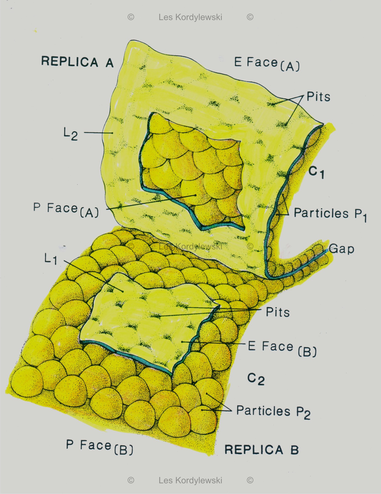
![[Figure 1]](/gapjunction/figures/Fig.%201.jpg)
![[Figure 2]](/gapjunction/figures/Fig.%202.jpg)
![[Figure 12]](/gapjunction/figures/Fig.%2012.jpg)
![[Figure 13]](/gapjunction/figures/Fig.%2013.jpg)
![[Figure 14]](/gapjunction/figures/Table%20I%20(Fig.%2014).jpg)
Gap Junction Ultrastructure Controversy Resolved in 3-D
Gap Junction Ultrastructure Controversy Resolved in 3-D: PITS AND PARTICLES DO NOT MATCH.Copyright © 2022 Les Kordylewski
Explanation:
The text of the attached article entitled: "Complementary replication of Gap Junctions in sheep cardiac Purkinje strands; Evidence for noncomplementarity of particles and pits” is the original manuscript describing the results of my observations of the three-dimensional structure of Gap Junctions (GJ) in the cell membranes of the myocytes of heart. Additionally, I am posting another never published before review article entitled: "Gap Junction ultrastructure controversy resolved in 3D. Pits and particles do not match." which explains in detail my methods of visualization details of the cell membrane ultrastructure in 3D.
The presented observations of GJ details after application of the freeze-fracture (FF) technique, were a by-product of my research on the ultrastructure of myocyte cell membranes, carried out between 1982 and 1995 in the Electron Microscopy Laboratory of the Department of Medicine of the University of Chicago. My results were the subject of a series of publications (American Journal of Physiology, 245 (6), H992-H997, 1983; Biophysical Journal, 47, 2, 2, 506a, 1985; American Journal of Physiology, 248 (3, Pt. 2), H297-H304, 1985; Developmental Biology, 112 (2), 485-488, 1985; Tissue and Cell, 18 (5), 793-801, 1986; Circulation Research, 73 (1), 135-146, 1993; Journal of Microscopy, 173 (3), 173-81, 1994; Journal of Histochemistry and Cytochemistry, 43 (5), 481-488, 1995), which are widely cited.
To better visualize particles in these membranes and to examine them quantitatively, I consistently used three-dimensional imaging (by taking hundreds of photographs using electron microscope equipped with a tilting table) and analyzing such stereopairs. I used the same method taking EM pictures of membrane areas with split GJ, while on the complementary replicas I found complementary Face E and P, previously adjacent to each other before splitting the same Gap Junction (GJ).
This manuscript has never been published. My observations and conclusions (demonstrating the lack of complementarity of pits and particles in the complementary replicas of split cell membranes, carried out on the three-dimensional images (stereopairs) of the same areas of GJs in Face E and P in the replicas of the same GJ) were the subject of crushing criticism, as being inconsistent with the commonly accepted basic model of the structure of GJ. This traditional model, in which pits are supposed to be scars in Face E after particles visible in Face P were pulled out of them in the FF process, is still presented in this way in every textbook of Cell Biology.
After reporting my results at the 1984 Cell Biology Conference in Baltimore (Are gap junctional pits and particles complementary structures? Biophysical Journal, 47, 2, 2, 506a, 1985), I was discouraged to publish the full text of this controversial article, because I was at risk of losing funding for my research. Therefore, I did not send this manuscript for publication, although I was (and I still am) convinced that my conclusions refuting the old model are correct.
In the examination of GJs no one has ever used my original comparative method of matching Face E and P pages by superimposing three-dimensional (3D) images of the replicas in the complementary P and E areas of the cell membrane interior of the same GJ. Usually, only the comparison of "flat" individual photos of face E and P was done, usually in different areas – not of the same GJ. Therefore, traditional conclusions were drawn that particles must always be complementary to pits in GJ. On the contrary, my observations of "matching" the details of complementary face E and P structures in replicas of membranes coming from the same areas of the same GJ led to the only possible interpretation that when three-dimensional images of Face E and P of the same Gap Junction were superimposed, the pits "fit" into the space between the connexons. This obviously contradicted the commonly accepted view that the pits are complementary to particles in the connexon, as if the pits were scars in Face E from P Face particles drawn from them. According to my observations, between the pits there are halves of connexons broken in the middle by FF. In the replicas they appear as complementary to the sister set of halves visible as particles on the Face P side.
At the time when I made this observation, I used numerous simple devices and techniques available to me for the evaluation of high-resolution black and white microphotographs – electron micrographs (see subsection Material and Methods). To confirm the controversial complementarity of the structures that I demonstrated, my excellent quality photographs would deserve to be processed now with more advanced contemporary computer methods. Unfortunately, at present I am not able to perform this task myself.
However, I consider this topic somewhat important, because it draws attention to the possible role of the space between the connexons, where in my opinion the pits are located. Despite the extensive literature on the connexons themselves, the importance of the space between them is underestimated. After all, just there the unknown forces that hold the connexons together are located. Nevertheless, the conclusions about the role and structure of the connexons are usually drawn through the use of theoretical models, and not as a result of directly revealing their structure. On the other hand, the factors which keep individual connexons together in a common assembly, are probably located in the spaces between the connexons (appearing as pits), and remain underestimated.
All Rights Reserved.